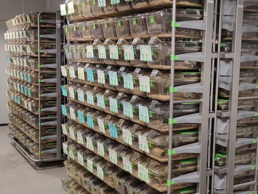
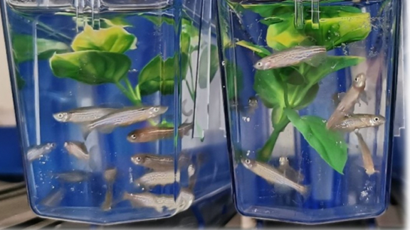
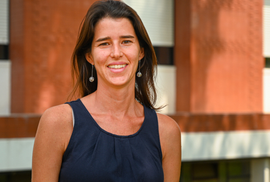
English | Português

Mitose

Interesse da Investigação

Cell-cycle regulation, centrosome duplication and separation, chromosome condensation, molecular motors, microtubules.

Alvaro Tavares

Ph.D. in Medical Science

Universidade de Porto, Porto


Investigador Principal
Telefone 21 440 7943
Exensão 243
Email
Local (Ala) Zheng Ho (C1) - Sala 1C

Membros do Grupo

Cláudia Florindo Postdoc
Tel: 21 440 7917


Projecto de Investigação

Regulation of proteolysis during mitosis

The ubiquitin-proteosome pathway is a major proteolytic system acting in various cellular processes (Hershko and Ciechanover 1998). In this system, the proteins are tagged with multi-ubiquitin chains and are then degraded by the 26S proteosome. The 26S proteosome is made up of two subcomplexes: the 20S proteosome and the regulatory complex. The regulatory complex consists of 18 subunits with molecular masses of 28-110kDa, including 6 putative ATPases (Rtp1-Rtp6) and 12 non-ATPase subunits (Rpn1-11) (Finley et al 1998, Holzl et al 2000). One level of control is obviously at the step of ubiquitination, and in fact most mutants described are components of the different ubiquitin-ligase complexes (eg. Glotzer and Dechant 2002). The 26S proteosome is believed to be constitutively active and has not attracted much attention as a regulatory molecule. However, according to recent progress in structural analysis of the proteosome the specificity of proteolsis pathway may well be modulated by the 26 proteosome. Phosphorylation of proteosome components C8 and C9 seems to increase proteolytic activity, but the responsible kinase has not been identified. In this project we study the effects of mutations in a gene coding for a component of the regulatory complex, the protein Rpn9. Surprisingly, we have found that mutations in this gene result only in abnormal mitosis, suggesting that it is required to selectively direct the degradation of proteins during this stage of the cell cycle. We wish to determine the nature of these proteins, and for such we will use a combined approach of RNAi and MALDI mass spectometry. In addition, the detailed characterization of the mutant phenotype, using classical genetics and confocal microscopy, will help to clarify the role of those proteins during mitosis (or better say, what happens if they’re not degraded) and of the proteosome itself.

Colaboradores

Hungary
Peter Deak

FCT/UNL
Rui Gomes

Projecto de Investigação

Role of mammalian Mob-like proteins in determining the timing of cytokinesis

The aim of this project is to study the cell division mechanism, in particular the aspects regulating the formation of a bipolar mitotic spindle. More specifically, we intent to study the human Mob1-like proteins (HsMobs). Our results indicate that HsMobs are centrosomal proteins essential for mitotic spindle assembly and for cytokinesis, and RNAi experiments resulted in an increase of multinucleated cells. The four different HsMob proteins, although co-localising at the centrosomes during mitosis, seem to have different functions. We which to precise the function of each HsMob protein and define if they are all essential for spindle assembly, cytokinesis, or both. For such, we are applying a concerted approach using molecular biology, protein crystalography, and mouse genetics, congregating three different groups. We explore the in vivo function of the HsMob in tissue culture cells by preventing the expression of each protein, using RNAi protocols. To this end we observe fixed or live cells by confocal microscopy, after transfection with dsRNA. We use time-lapse microscopy and serial section EM to define the in vivo behavior of centrosomes during the vertebrate somatic cell cycle, expecting this way to define the role of each HsMob during the differents phases of the cell cycle. Time-lapse will also be used to caracterise the dynamic intracelular localization of each HsMob, tagged with GFP- or CFP-proteins. Finally, interacting proteins will be identified by phage-display. In order to analyse the function of this class of proteins throughout development, in particular if their supression leads to tumour formation, we will generate conditional knockout mice. We will use the Cre-recombinase loxP system in order to be avoid possible early lethal phenotypes, and to be capable of determining the gene function in specific tissues. Another important objective is to determine the three-dimensional structure of the Mob1-like proteins by X-ray diffraction. Few centrosomal proteins have been crystalised to date and we hope this way, together with the information gathered in the other tasks, to be capable of establishing a relationship struture-function for these centrosomal proteins. This will allow the design of new molecules capable of interfering with HsMob function with potential therapeutic applications, namely prevent cell proliferation. The identification of HsMob molecular partners can also give important clues to about cancer progression.

Colaboradores

ITQB/UNL
Maria Arménia Carrondo

Cambridge, UK
Jon Pines

Projecto de Investigação

Mitotic roles for the Plkk and Mps1 kinases

In a typical somatic cell cycle, M-phase comprises mitosis and cytokinesis. M-phase progression is largely controlled by protein phosphorylation, and several protein kinase families have already been implicated in such control. The aim of this project is to study the cell division mechanism, more specifically, we intend to study the function of the proteins kinases DPlkk and DMps1. The kinase Plkk was originally is Xenopus described as the activator of the mitotic kinase polo, and Mps1 protein is an essential S. cereviseae kinase involved in the mitotic checkpoint and in the duplication of the spindle pole body. In preliminary work conducing to this project we have cloned the Drosophila homologues of the kinases Plkk and Mps1. We will address the function of these kinases following a molecular biology and genetics concerted approach. We already have a DPlkk mutant allele created by insertion of a P-transposable element into the gene. The mutant is lethal in the early embryos stages, much like strong alleles of polo. As there is no mutant alleles of DMps1 we will analyse, in Drosophila S2 cells, the effects of a lack of function brought about by transfection with double-stranded RNA (dsRNA) although the constratuction of such a mutant is simultaneously being done. One of the major breakthroughs we expect with this project is the finding of physiological kinase substrates. We will look for substrates of the DPlkk and DMps1 in preparations of centrosomes and study how phosphorylation by these kinases can modify the function of the substrates. Very powerful in vitro systems have been developed in Drosophila extracts that allow biochemical and biophysical approaches to the function of the centrosome (eg. Carmo Avides and Glover 1999). We will utilise this in vitro system to study the roles of both kinases in centrosomal microtubule nucleation (as done for the kinase polo, Carmo Avides et al 2001). We will also look for DPlkk and DMps1 interacting proteins, using affinity columns to isolate protein complexes capable of binding these proteins. Our approach will be complemented by database searches for human ESTs to identify the human orthologues of novel proteins identified in Drosophila. We hope with these different approaches to advance the discovery of new centrosome and spindle proteins and elucidate their function in the regulation of chromosome segregation.

Projecto de Investigação

Molecular and biochemical analysis of centrosome components in Drosophila melanogaster

The accurate segregation of chromosomes at mitosis is essential for the provision of genetic material to ensure cell viability. Defects in any stage of this process can lead to cell death or, in higher organisms, the development of cancer. Multipolar spindles have often been observed in human cancers in situ as well as an abnormal number of centrosomes. Identification of the molecular targets of centrosome kinases and elucidation of the pathways that regulate centrosome duplication, separation and function provide novel opportunities for therapeutic intervention. We previously showed that the protein kinase polo is a centrosomal kinase, and that is required for the formation of a bipolar spindle and for the proper execution of cytokinesis. We wish to understand how the activity of the polo protein kinase is regulated and how it functions at the level of the centrosomes. We previously found that polo proteins, either from Drosophila embryo extracts or from Xenopus egg extracts, bind to several proteins forming different stable complexes. We are now on the process of identifying the complexe's components in total embryo extracts and in centrosome preparations. We want to characterize these proteins, sorting which are polo substrates and which are activators. Taking advantage of Drosophila genetics we have also searched and isolated new genes required for spindle assembly and centrosome function, some of which coding for proteins with high degree of homology with the Saccharomyces cereviseae proteins. We are now on the process of characterising these genes.

Colaboradores

Cambridge, UK
David Glover

Publicações

Adams, R., Tavares, A., Salzberg, A., Bellen, H. e Glover, D. (1998). pavarotti encodes a kinesin-like protein required to organise the central spindle and contractile ring for cytokinesis. Genes & Dev. 12 :1483-1494

Carmena, M., Riparbelli, M., Minestrini, G., Tavares, A., Adams, R., Callaini, G. e Glover, D. (1998). Drosophila polo kinase is required for cytokinesis. J. Cell Biol 143 :659-671

Wianny, F., Tavares, A., Evans, M., Glover, D. and Zernicka-Goetz, D. (1998). Mouse polo-like kinase 1 associates with the acentriolar spindle poles, meiotic chromosomes and spindle midzone during oocyte maturation Chromosoma 107 :430-439

Glover, D., Hagan, I. and Tavares, A. (1998). Polo-like kinases: a team that plays throughout mitosis Genes & Dev. 12 :3777-3787

Tavares, A., Glover, D. e Sunkel, C. (1996). The conserved mitotic kinase polo is regulated by phosphorylation and has preferred microtubule-associated substrates in Drosophila embryo extracts. EMBO J. 15 :4873-4883